
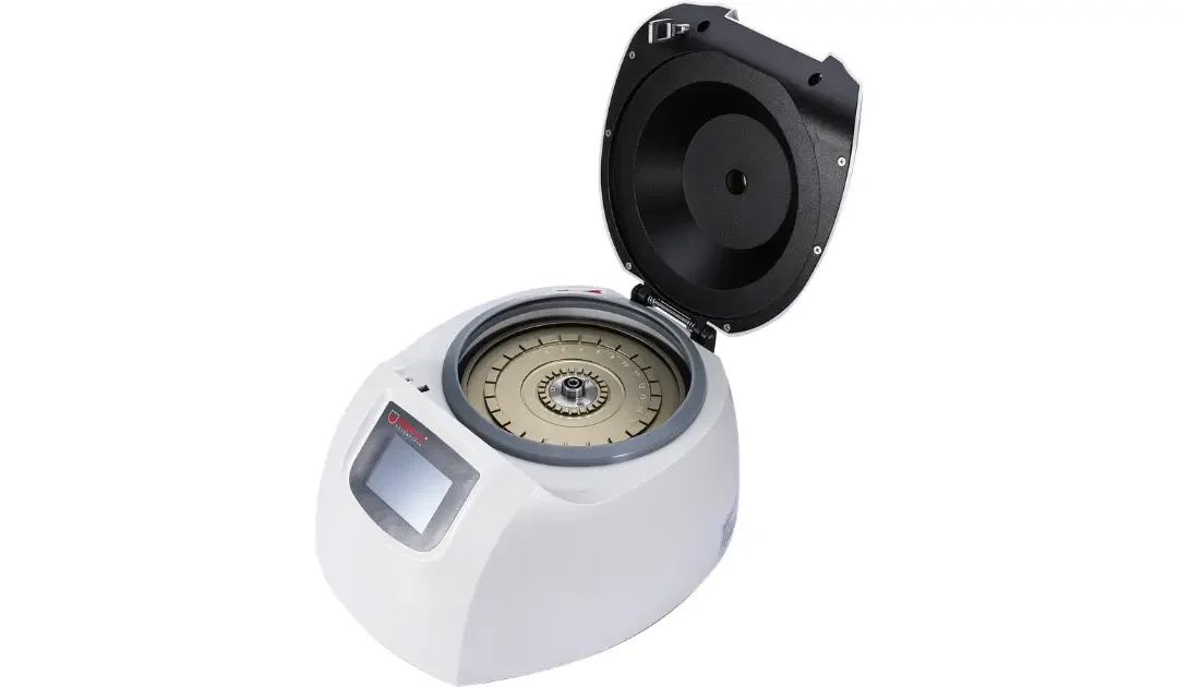
The hematocrit centrifuge is one of the most clinically essential instruments in a diagnostic laboratory setting, yet it is also one of the simplest in concept. It spins small capillary tubes filled with blood at high speeds, separating the blood into its component layers so that the proportion of red blood cells relative to total blood volume can be precisely measured. That measurement, the packed cell volume or PCV, is one of the most informative and straightforward diagnostic values in clinical hematology.
This guide covers the working principle of the hematocrit centrifuge, its clinical applications, how to interpret results, and what to look for in the instrument itself.
What The Hematocrit Value Measures
The hematocrit, also referred to as packed cell volume (PCV) or erythrocyte volume fraction (EVF), represents the proportion of blood volume occupied by red blood cells. According to the National Institutes of Health StatPearls documentation on hematocrit, normal hematocrit values are approximately 48 percent for men and 38 percent for women, though values vary with age, sex, and altitude.
This single percentage value provides clinically significant information about a patient’s oxygen-carrying capacity, hydration status, and overall blood health. The measurement is considered an integral part of a complete blood count (CBC), alongside hemoglobin concentration, white blood cell count, and platelet count.
How A Hematocrit Centrifuge Works
A hematocrit centrifuge spins capillary tubes containing blood at speeds typically between 10,000 and 12,000 RPM for three to five minutes. The centrifugal force separates blood into three distinct layers according to density. At the bottom, the heaviest component, red blood cells, pack tightly together. Above them lies a thin buffy coat layer of white blood cells and platelets. Above the buffy coat is the plasma, the least dense component, which remains at the top.
After centrifugation, the packed cell volume is read using a microhematocrit reader card or a built-in digital reader. The PCV is calculated as the proportion of the total tube length occupied by the packed red blood cell layer, expressed as a percentage. This measurement is considered the most accurate and simplest of all tests in clinical hematology for detecting the presence and degree of anemia or polycythemia.
Primary Hematocrit Centrifuge Uses In Clinical Diagnostics.
Anemia diagnosis and monitoring: A hematocrit below the normal range indicates fewer red blood cells than expected, which reduces the blood’s oxygen-carrying capacity. The hematocrit centrifuge provides the PCV value that, combined with other CBC parameters, allows clinicians to diagnose anemia, classify its type (iron deficiency, hemolytic, aplastic), and monitor the response to treatment over time.
Polycythemia detection: Conversely, an elevated hematocrit indicates polycythemia, a condition where the red blood cell mass is excessive. This can cause increased blood viscosity, elevated clotting risk, and cardiovascular complications. NCBI Bookshelf documentation confirms that hematocrit measurement is a simple test used to identify conditions such as polycythemia and to monitor the response to treatments.
Dehydration assessment: Dehydration reduces plasma volume without proportionally reducing red blood cell mass, causing a relative increase in hematocrit. Tracking hematocrit in patients with suspected dehydration or during rehydration therapy provides objective evidence of fluid status changes.
Pre-surgical screening: Baseline hematocrit is routinely measured before surgical procedures to assess whether a patient has adequate red blood cell reserves to tolerate potential blood loss during the operation.
Veterinary and animal research applications: Hematocrit centrifuges are used in veterinary diagnostics and laboratory animal research for routine hematological analysis of small blood volumes from companion animals, laboratory rodents, and other research subjects. The small capillary tube format is well-suited for the limited blood volumes available from small animals.
Reading And Interpreting Hematocrit Results
After centrifugation, the packed red blood cell layer is measured using a microhematocrit chart or reader. The length of the red cell layer is divided by the total blood column length and multiplied by 100 to express the result as a percentage. A result below the reference range for the patient’s sex and age is reported alongside the clinical context: low hematocrit combined with low hemoglobin and small red cell size suggests iron deficiency anemia; low hematocrit with large red cells suggests macrocytic anemia. Normal values for newborns range from 55 to 68 percent according to clinical physiology documentation.
The Dm2412 Hematocrit Centrifuge
The DM2412 Hematocrit Centrifuge from NE LabSystems accommodates 24 capillary tubes simultaneously and operates at speeds up to 12,000 RPM, generating the centrifugal force required for accurate PCV determination. It features digital controls, programmable timing, and a built-in reading scale for direct hematocrit measurement post-spin. Backed by a free extended warranty on U.S. purchases and factory-trained engineering support, it is a reliable choice for clinical diagnostic labs running routine hematocrit testing. Call (877) 733-6838 for more information.